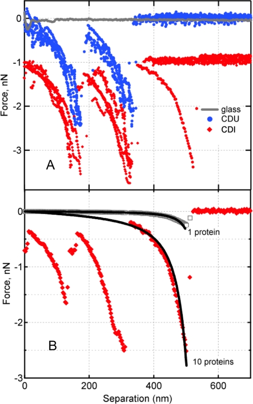
FIGURE 3.
A, representative force spectra between Fn and putative FnBP expressed on the external envelop of S. aureus isolates from patients with an infected cardiac device (CDI, red diamonds) or the bloodstream of patients with an uninfected cardiac device (CDU, blue circles). Nonspecific adhesion (attractive interaction at separation ∼0 nm) (68) between Fn and a glass surface is shown in gray. By convention, negative values indicate attractive force in nanonewtons (10−9 N). The CDI traces are offset vertically to ease visual comparison. B, the worm-like chain model (Equation 1) was used to describe the force-extension profile for 1 protein or 10 parallel proteins (black curves). Following the protocol of Refs. 16 and 17, the 10-bond scenario was calculated as the summation of 10 individual polypeptide chains. Also shown are two experimentally measured protein extension traces preceding the specific bond rupture events. These show the rupture of one bond, observed rarely for Fn tip on clean glass (gray), versus 10 Fn–FnBP bonds, commonly observed for Fn tip on S. aureus (red).

