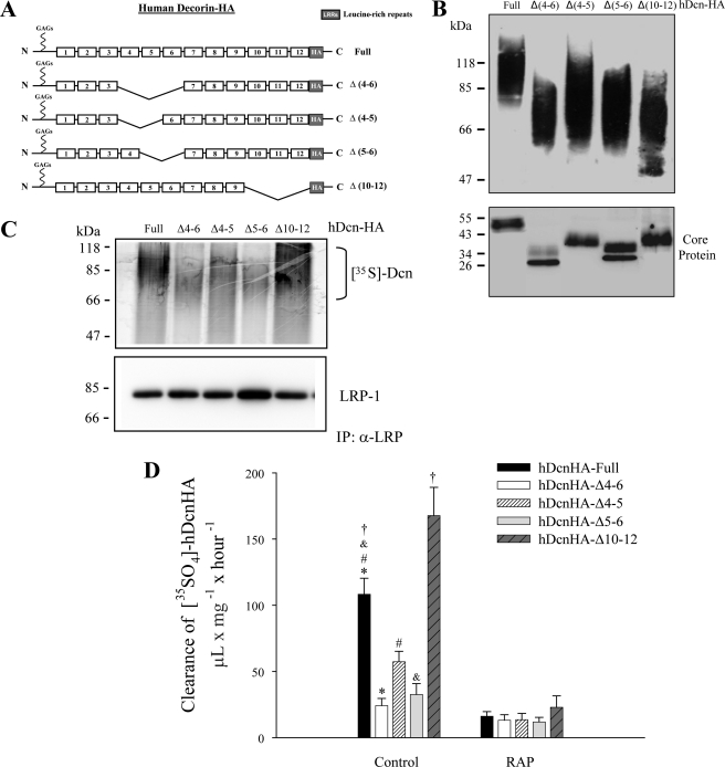
FIGURE 1.
The LRR4–6 decorin region is necessary for LRP-1 binding and LRP-1-mediated decorin endocytosis. A, schematic showing decorin deletion mutants. B, deletion mutants generated in CHO-K1, which lack LRR4 and 5 (Δ4–5); 4, 5, and 6 (Δ4–6); 5 and 6 (Δ5–6); or 10, 11, and 12 (Δ10–12); were purified by DEAE-Sephacel and separated in a 4–12% SDS-PAGE gradient for detection of glycanated forms (upper) or protein-core after treatment with chondroitinase ABC (lower). Decorins were visualized by Western blot using anti-HA antibodies. C, C2C12 cells were incubated with 35S-labeled decorin mutants at 4 °C for 3 h. Extracts were immunoprecipitated with anti-LRP-1 antibodies and the presence of decorin deletion mutants in the immunoprecipitate (IP) was evaluated by autoradiography (18). Protein levels were detected in the immunoprecipitate with an anti-LRP-1 antibody by Western blot. D, C2C12 myoblasts were incubated with 35S-labeled decorin mutants at 37 °C for 3 h in the absence (control) or presence of 1 μm receptor-associated protein (RAP), then cells were analyzed to determine the level of endocytosis as described before (5). Values correspond to the mean ± S.D. from three independent experiments (*, #, and &, p < 0.001; †, p < 0.05).

