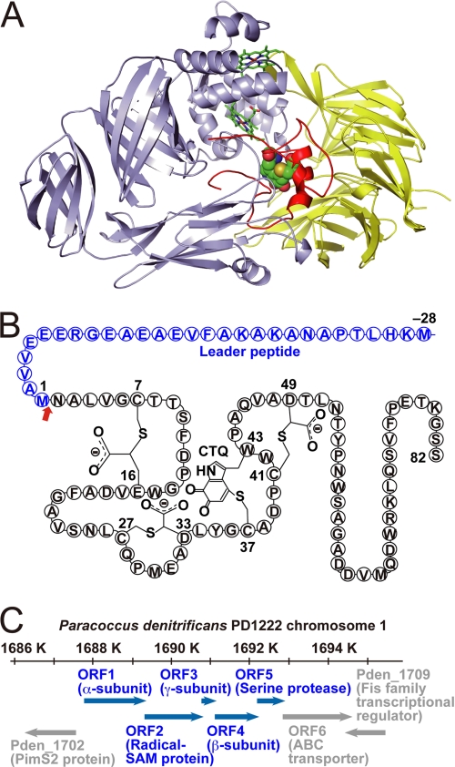
FIGURE 1.
Protein and gene structure of QHNDH. A, ribbon model of QHNDH from P. denitrificans (PDB code 1JJU). The light blue, yellow, and red ribbons represent the α, β, and γ subunits, respectively. The two hemes in the α subunit are indicated as sticks, and CTQ atoms in the γ subunit are depicted as balls. B, schematic of the γ subunit polypeptide with the 28-residue N-terminal leader peptide and intrapeptidyl modifications (three Cys-to-Asp/Glu thioether cross-links and CTQ) at the indicated residue numbers. The red arrow indicates the cleavage site of the leader peptide. C, schematic of the gene structure of QHNDH in chromosome 1 of Pd1222, ORF1-ORF5, and the peripheral genes are shown as blue and gray arrows, respectively. Gene identification numbers from the NCBI genome database are included, with the annotated proteins referred to in parentheses. SAM, AdoMet.

