FIGURE 3.
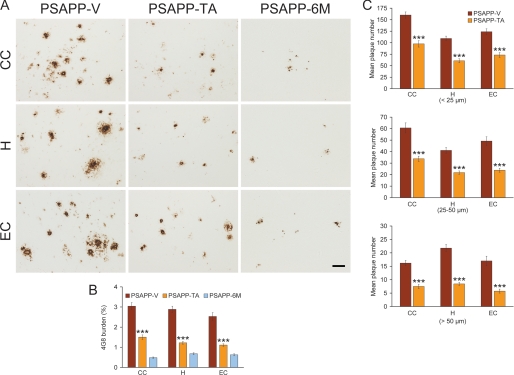
Cerebral parenchymal β-amyloid deposits are mitigated in tannic acid-treated PSAPP mice. Data were obtained from PSAPP mice treated with vehicle (PSAPP-V, n = 16) or TA (PSAPP-TA, n = 16) for 6 months commencing at 6 months of age (mouse age = 12 months at sacrifice). To examine if the TA treatment delayed versus prevented disease progression, untreated PSAPP mice (PSAPP-6M, n = 12) at 6 months of age were also included in these analyses. A, representative photomicrographs of 4G8 immunohistochemistry are shown for cerebral β-amyloid plaques in PSAPP-V, PSAPP-TA, and PSAPP-6M mice. Brain regions shown include: cingulate cortex (CC, top), hippocampus (H, middle), and entorhinal cortex (EC, bottom). Scale bar denotes 50 μm. B, quantitative image analysis for Aβ (4G8) burden is shown, and each brain region is indicated on the x axis. C, morphometric analysis of cerebral parencymal β-amyloid plaques is shown in PSAPP-V and PSAPP-TA mice. Brain sections were stained with 4G8 antibody, and plaques were counted based on maximum diameter and assigned to one of three mutually exclusive categories: small (<25 μm; top), medium (between 25 and 50 μm; middle), or large (>50 μm; bottom). Mean plaque subset number per mouse is shown on the y axis, and each brain region is represented on the x axis. The statistical comparison for B is within the brain region and versus PSAPP-TA mice, and for C, between PSAPP-V and PSAPP-TA mice.

