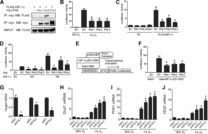
FIGURE 3.
All three FHL proteins inhibit HIF-1 transcriptional activity. A, FHL2 is the only FHL family member that binds HIF-1α. 293T cells were co-transfected with FLAG-HIF-1α and either EV or vector encoding Myc-FHL1, Myc-FHL2, or Myc-FHL3. Lysates were subjected to immunoprecipitation (IP) with anti-Myc antibody and probed for FLAG-HIF-1α and Myc-FHL by WB. B, FHL1–3 inhibit HIF-1 activity induced by hypoxia. 293 cells were co-transfected with p2.1, pSV-RL, and EV or vector encoding FHL1, FHL2, or FHL3. At 24 h post-transfection, cells were exposed to either 20% O2 or 1% O2 as indicated for another 24 h, then lysed and the ratio of firefly:Renilla activity was determined. (C) FHL1–3 inhibit HIF-1 activity induced by HIF-1α overexpression under nonhypoxic conditions. 293 cells were co-transfected with p2.1, pSV-RL, FLAG-HIF-1α vector or EV, and either EV, FHL1, FHL2, or FHL3 vector. 24 h post-transfection, the cells were lysed, and the ratio of firefly:Renilla activity was determined. D, FHL1–3 inhibit HIF-1 in a hydroxylation-independent manner. 293 cells were co-transfected with pSV-RL; p2.1; vector encoding wild-type FLAG-HIF-1α (WT) or P402A/P564A/N803A triple mutant (TM); and either EV, FHL1, FHL2, or FHL3 vector. 24 h post-transfection, the cells were lysed to determine firefly:Renilla activity. E, schematic of HIF-1α TAD assay. F, FHL1–3 inhibit HIF-1α TAD function. 293 cells were co-transfected with pSV-RL; pG5-E1b-Luc; either Gal4-HIF-1α(531–826) or Gal4-EV; and either EV, FHL1, FHL2, or FHL3 vector. 24 h post-transfection, the cells were exposed to hypoxia for an additional 24 h. The cells were harvested, and the ratio of firefly:Renilla luciferase was determined. The amount of total plasmid transfected was kept constant in all experiments. The results are shown as the means ± S.D. G, HeLa cells were transfected with either empty shRNA vector (shEV) or shRNA targeting FHL1, FHL2, or FHL3. 48 h post-transfection RNA was isolated, and quantitative RT-PCR of the target mRNA was performed. H–J, HeLa cells were transfected with either shEV or shRNA targeting FHL1, FHL2, or FHL3. 24 h post-transfection the cells were exposed to either 20% O2 or 1% O2 for an additional 24 h. RNA was isolated, and quantitative RT-PCR was performed against GLUT1 (H), PDK1 (I), and VEGF (J). The results are shown as the means ± S.E. *, p < 0.01 compared with EV or shEV.

