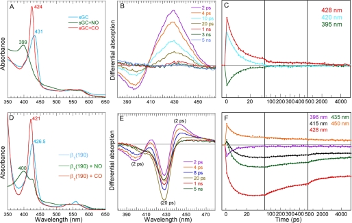
FIGURE 1.
Dynamics of NO after photo-dissociation from the entire sGC and from the isolated heme domain β1(190). A, steady-state spectra of unliganded sGC and liganded with NO and CO. B, transient spectra at selected time delays after photo-dissociation of NO from full-length sGC. C, kinetics of NO rebinding to sGC at selected wavelengths. D, steady-state spectra of unliganded isolated heme domain β1(190) and liganded with NO and CO. The slope below 360 nm is due to the absorption of dithionite (0.5 mm) used as reductant. E, transient spectra at selected time delays after photo-dissociation of NO from β1(190). F, kinetics of NO rebinding to β1(190) at selected wavelengths fitted to a sum of exponentials. The averaged fitted parameters are given in Table 1 (Global SVD analysis in supplemental Fig. S1 and individual parameters in supplemental Table S1).

