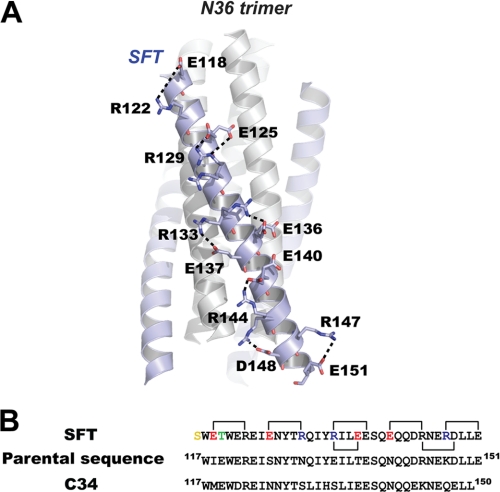
FIGURE 2.
Salt bridges formed by charged residues on SFT. A, ribbon model of 6-HB formed by SFT/N36 with the charged residues involving ion pair formation on the SFT helix shown as a stick model with the labels. The salt bridges formed between charged residues are indicated with dashed lines. B, sequence alignment of SFT, its parental sequence (HIV-1 subtype E gp41 CHR(117–151)), and C34. Numbering is based on the sequence of HXB2 gp41. The color codes for SFT sequence are as follows: yellow, N-terminal capping acetylserine; green, mutation to stabilize the hydrophobic pocket; red, residues were changed to negatively charged residues; blue, residues were changed to positively charged residues; black, unchanged residues. Salt bridges observed in the crystal structure of SFT are indicated with the linkers.

