FIGURE 5.
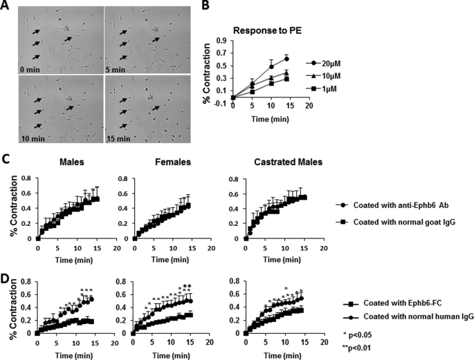
Reverse but not forward signaling between Ephb6 and Efnbs dampens VSMC contractility. Data presentation and statistical analysis are the same as described for Fig. 3 (C and D). A, micrographs of VSMC contraction after PE stimulation. VSMC were stimulated with 20 μm PE and imaged every min. Images at 0, 5, 10, and 15 min are presented. Four arrows point to the same four cells during the 15-min imaging period and show their contraction. The photos also reveal that ∼85% of the cells are capable of contraction, indicating the purity of VSMC in such cell preparations. B, dose-dependent response of VSMC contractility to PE stimulation. WT VSMC were stimulated with PE at different concentrations for 15 min at 37 °C. C, solid phase anti-Ephb6 Ab had no effect on VSMC contractility. Wells were coated with goat anti-Ephb6 Ab or normal goat IgG (2 μg/ml during coating). VSMC from WT male (left panel), WT female (middle panel), or castrated WT male (right panel) mice were cultured in these wells for 4 days and then stimulated with 20 μm PE. D, solid phase Ephb6-Fc reduced VSMC contractility. The wells were coated with recombinant Ephb6-Fc or normal human IgG (2 μg/ml during coating). VSMC from WT male (left panel), WT female (middle panel), or castrated WT mice (right panel) were cultured in these wells for 4–5 days and then stimulated with 20 μm PE.

