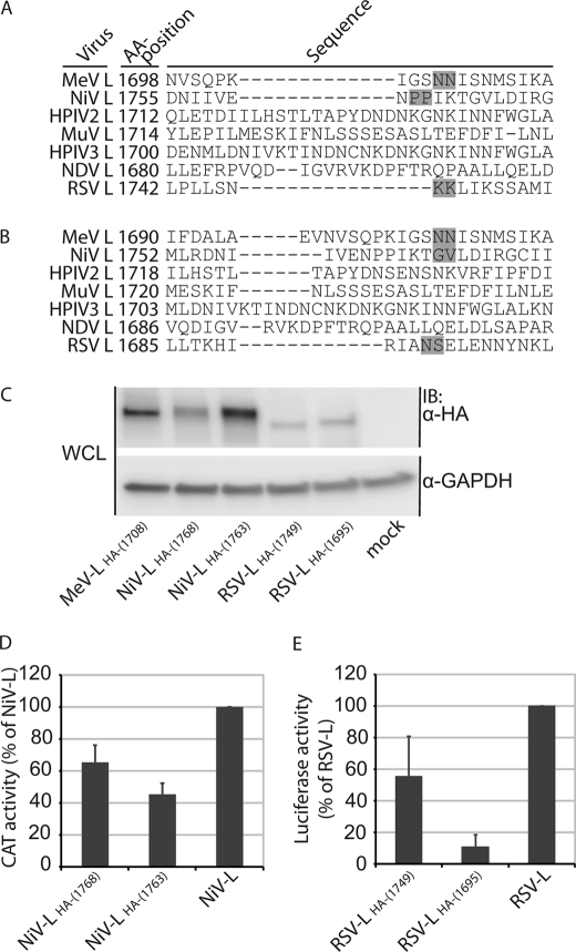
FIGURE 4.
Identification of HA epitope tag insertion sites in NiV and RSV L. A and B, sequence alignments of L proteins derived from MeV-Edm, NiV (88), HPIV2 (89), MuV-Miyahara (90), HPIV3–14702 (91), NDV-AF2240 (80), and RSV-A2 using the ClustalW2 (A) and MUSCLE (B) algorithms are shown. Regions corresponding to part of the LR II/LR III intersection in MeV L are shown, and numbers reflect amino acid positions. Gray boxes mark the insertion sites of the epitope tags. C, shown is expression analysis of the newly generated NiV and RSV L variants. Whole cell lysates (WCL) of BSR-T7/5 cells transfected with the different L expression plasmids were gel fractionated and immunostained (IB) using specific antibodies directed against the HA epitope or cellular GAPDH. Control cells (mock) received empty vector in place of L-encoding plasmid. D and E, shown is activity testing of the HA-tagged NiV and RSV L variants using specific replicon reporter systems for NiV (D, CAT reporter) and RSV (E, firefly luciferase reporter). Values are expressed relative to activities of standard NiV or RSV L and reflect the averages of four independent experiments ± S.D.

