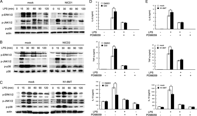
FIGURE 4.
Notch signal inhibits TLR-triggered inflammatory cytokine production by ERK1/2 inactivation. A–C, mouse primary peritoneal macrophages were transfected with NICD1 (A), NICD2 (B), N1-6MT (C), or mock vector. After 48 h, cells were stimulated with 100 ng/ml LPS for the indicated time. Phospho-ERK, JNK, and p38 were detected by immunoblotting. Data are representative of three separate experiments. Similar results were obtained in three independent experiments. D, primary peritoneal macrophages were pretreated with 2 μm GSI or dimethyl sulfoxide (DMSO) for 24 h and stimulated with 100 ng/ml LPS and 10 nm PD98059 together or alone for 8 h. The supernatant IL-6, TNF-α, and IL-10 were measured by ELISA. E, mouse primary peritoneal macrophages were transiently transfected with N1-6MT or mock vector. After 48 h, cells were stimulated with 100 ng/ml LPS and 10 nm PD98059 together or alone for 8 h. IL-6, TNF-α, and IL-10 in the supernatants were measured by ELISA. Data are shown as mean ± S.D. (error bars) of three independent experiments. *, p < 0.05; **, p < 0.01.

