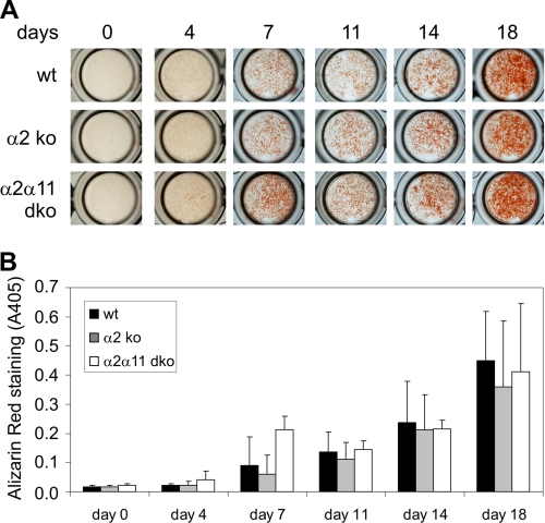
FIGURE 3.
Functional analysis of primary osteoblasts. Osteoblasts were isolated from calvaria of newborn wild-type (wt) mice and those deficient in α2β1 (α2 ko) or double-deficient (α2α11 dko) and cultured for up to 14 days with and without differentiation medium containing 2-phosphoascorbic acid and β-phosphoglycerate. A, osteoblast cultures were stained with Alizarin red at various time points. B, amounts of incorporated dye (mean ± S.D.) reflecting the degree of mineralization, quantified by assessing absorption at 405 nm, revealed no obvious differences between genotypes. n ≥ 5 animals (= cultures) per genotype. Each culture was analyzed in triplicate wells per assay.

