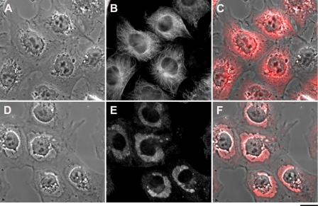
FIGURE 5.
Effect of bufalin on tubulin localization. The experiments were conducted as described in the legend to Fig. 1. Immunostaining of tubulin was performed on NT2 cells grown in DMEM-F12 on glass coverslips for 24 h. The DMEM-F12 was then replaced with medium with (D–F) or without (A–C) 20 nm bufalin for 4.5 h. The cells were fixed with 1.5% glutaraldehyde and stained with anti α-tubulin monoclonal antibodies, and images were acquired. A and D, phase microscopy; B and E, fluorescence microscopy; C and F, merged phase-contrast and fluorescence. Scale bar, 20 μm.

