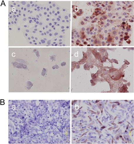
FIGURE 2.
Immunocytochemistry for HMOCC-1 of RMG-I cells and CHO Lec2 cells. Cells were treated with the HMOCC-1 antibody (human IgM) and stained using the immunoperoxidase method. Hematoxylin was employed as a counterstain. A, immunocytochemistry of RMG-I cells before (panels a and b) and after (panels c and d) mild acid hydrolysis with (panels b and d) or without (panels a and c) HMOCC-1 antibody. B, CHO Lec2 cells transfected with mock vector (panel a) or co-transfected with GAL3ST3 plus B3GNT7 expression vectors (panel b).

