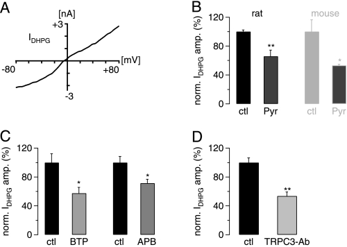
FIGURE 1.
The mGluR1-mediated slow EPSC in juvenile rat cerebellar slices is TRPC3-dependent. A, shown is the current-voltage relationship of the DHPG-induced inward current (IDHPG) (20 μm DHPG in P15 rat). B, DHPG-mediated inward current (IDHPG) (50 μm DHPG) was significantly blocked in the presence of 10 μm Pyr3 (Pyr) both in juvenile rat (P12–14; left pair of black bars; n = 5 for control (ctl) and 6 for Pyr3) and in juvenile mice (P12–14; right pair of gray bars; n = 3 for both control and Pyr3). IDHPG amplitudes in the presence of Pyr3 were normalized (norm.) to average control amplitude in absence of Pyr3. There was no significant difference in the extent of Pyr3-mediated block between rat and mouse (p = 0.3484; unpaired Student's t test). Preincubation in Pyr3 was 40 min. C, DHPG-mediated inward current (IDHPG) (50 μm DHPG) was significantly blocked by 15 μm BTP2 (left two columns; n = 4 for control (ctl) and 6 for BTP2 (BTP)) and by 10 μm 2-APB (APB; right two columns; n = 3 for each control and test; p = 0.0481, unpaired Student's t test). D, DHPG-mediated inward current (IDHPG) (50 μm DHPG) was significantly blocked by intracellular TRPC3-antibody (TRPC3-Ab); n = 5 for control (ctl) and 4 antibody experiments. IDHPG amplitudes in the presence of TRPC3 antibody were normalized (norm.) to average control amplitude in the absence of the antibody. Intracellular solution was supplemented with 4 μg/ml TRPC3 antibody. Reduction in current amplitude is significant (p = 0.0013; unpaired Student's t test).

