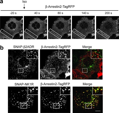
Figure 3.
Live cell imaging of HEK293A cells stably expressing SNAP-β2ADR or SNAP-NK1R transfected with β-arrestin2-TagRFP. From Supplementary Videos 3 and 4. (a) Images show the redistribution of β-arrestin2-TagRFP from the cytosol to the plasma membrane in the presence of isoproterenol in cells expressing SNAP-β2ADR. Boxes highlight the redistribution to the cell surface in adjacent cells. (b) Comparison of the colocalization of SNAP-β2ADR or SNAP-NK1R with β-arrestin2-TagRFP after agonist-induced uptake (isoproterenol for SNAP-β2ADR or Substance P for SNAP-NK1R) and 5 mM TCEP treatment. β-Arrestin2-TagRFP colocalizes with SNAP-NK1R labeled endosomes but not with SNAP-β2ADR. Scale bar, 10 μm.

