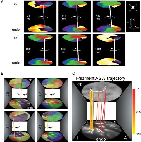
Figure 4.
I-filament ASWs in a PtAF heart. (A) Representative sequential colour phase maps of a reconstructed ASW spanning the anterior LAA wall. The white line joining the epicardial and endocardial singularity points indicates the presence of a sustained ASW whose I-shaped filament rotates clockwise during 1120 ms. (B) Meandering trajectory of an ASW filament during one rotation lasting 140 ms. Sequential colour phase maps and corresponding filament locations superimposed with matching real images of the endocardial and epicardial surfaces. (C) Tracing the filament trajectory. From 20 to 64 ms, the filament remained anchored to in thin myocardium bordered by thick pectinate muscles. Between 64 and 140 ms, the filament drifted across a pectinate muscle segment before anchoring to a neighbouring island of thin myocardium. Phase map colours as in Figure 1.

