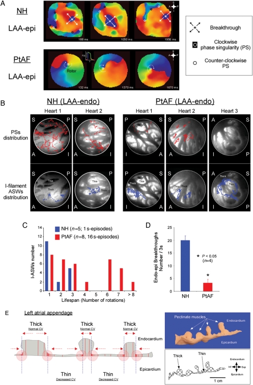
Figure 5.
AF dynamics in normal and PtAF hearts. (A) Representative sequential LAA-epi phase maps obtained from NH and PtAF hearts over a time window ∼1 s. In the NH (top), the dynamics were characterized by BTs and multiple PSs of both chiralities. In the PtAF heart (bottom), a sustained ASW was seen rotating clockwise throughout the recording period. Phase movies colours as in Figure 1. Inset; key for the various dynamic components. (B) Anatomical distribution of transient PSs (red dots, top) and I-filament ASWs lasting >3 rotations (blue dots, bottom) on the LAA-endo in two NH and three PtAF hearts. The PSs were transient in NH. In PtAF hearts, most PSs evolved into I-filament ASWs which meandered within the thin atrial myocardium or around thin-to-thick interfaces. S, superior; I, inferior; A, anterior; P, posterior. (C) Histogram of I-ASW's rotation numbers at the LAA in NH and PtAF. (D) Average number of BTs in NH and PtAF hearts. (E) Heterogeneous walls of LAA are substrates for scroll wave meandering. See text for details.

