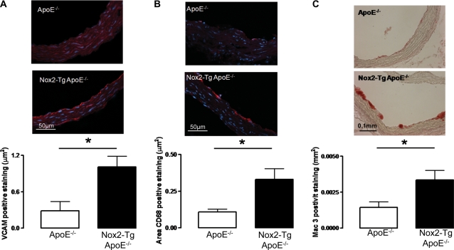
Figure 4.
Quantification of aortic root VCAM-1 and macrophage levels in Nox2-Tg ApoE−/− and ApoE−/− mice at 9 weeks of age. (A) Aortic root VCAM-1 staining (VCAM-1 positive cells stain red) was significantly greater in aortic roots from Nox2-Tg ApoE−/− mice compared with ApoE−/− littermates (P< 0.05, n= 5). (B) Aortic root macrophage content was quantified by immunohistochemical staining in frozen sections for CD68 (macrophages stained red); Nox2-Tg ApoE−/− mice had significantly greater macrophage content compared with ApoE−/− littermates (P< 0.05, n= 5–8). (C) Aortic root macrophage content was quantified by immunohistochemical staining in paraffin sections for Mac-3 (macrophages stained red); Nox2-Tg ApoE−/− mice had significantly greater macrophage recruitment compared with ApoE−/− littermates (P< 0.05, n= 5–8). For quantification of VCAM-1 and CD68 staining, the area of positive staining was normalized to vessel length.

