Figure 1.

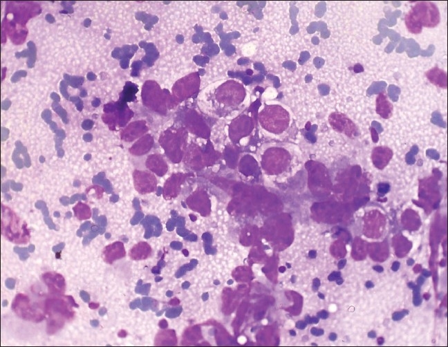
Photomicrograph of yolk sac tumor composed of irregular, large, cohesive three-dimensional cell balls and papillae, with infrequent single cells with abundant, vacuolated cytoplasm. (Leishman Giemsa, ×400)

Photomicrograph of yolk sac tumor composed of irregular, large, cohesive three-dimensional cell balls and papillae, with infrequent single cells with abundant, vacuolated cytoplasm. (Leishman Giemsa, ×400)