Figure 1.

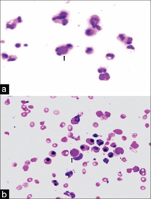
(a) Cytospin preparation of the pleural fluid showing plenty of lupus erythematosus (LE) cells (MGG, ×400). (b) Cytospin preparation of the pleural fluid showing tart cell (MGG, ×400)

(a) Cytospin preparation of the pleural fluid showing plenty of lupus erythematosus (LE) cells (MGG, ×400). (b) Cytospin preparation of the pleural fluid showing tart cell (MGG, ×400)