Figure 1.

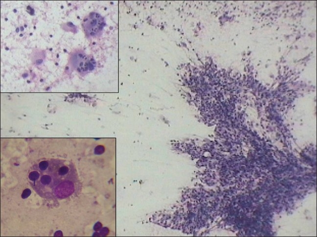
Cellular smears with clusters of histiocytes over a lymphocytic background (H and E, ×100). Upper inset shows histiocytes with emperipolesis of neutrophils (H and E, ×400) and lower inset shows emperipolesis of lymphocytes (Giemsa, ×1000)

Cellular smears with clusters of histiocytes over a lymphocytic background (H and E, ×100). Upper inset shows histiocytes with emperipolesis of neutrophils (H and E, ×400) and lower inset shows emperipolesis of lymphocytes (Giemsa, ×1000)