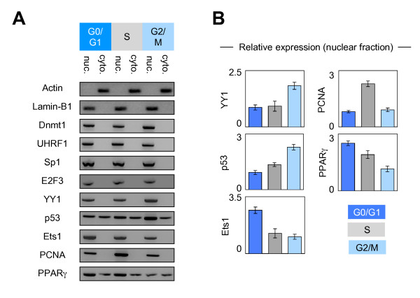
Figure 3.
Dynamic of expression and subcellular localization of the Dnmt1 and its partners of interaction during the cell cycle. (A) Pictures illustrate the results obtained after Western blot analyses. Nuclear (nuc.) and cytosolic (cyto.) were obtained by using the Nuclear Extract Kit (Active Motif, France). 30 μg and 50 μg of nuclear and cytosolic extracts were used for the western blot analysis concerning the Dnmt1, UHRF1, Sp1, E2F3, YY1, p53, Ets1, PCNA and PPARγ proteins. 20 μg of nuclear and cytosolic extracts were used for the western blot analysis concerning the actin and lamina-B1 proteins. (B) Graphs illustrate the relative expression of indicated proteins in nuclear fraction i.e. the results obtained from the densitometric analyses of western blot (Fusion Fx7 Imager, Fisher Scientific, France and ImageJ software). Lamina-B1 was used as internal control.

