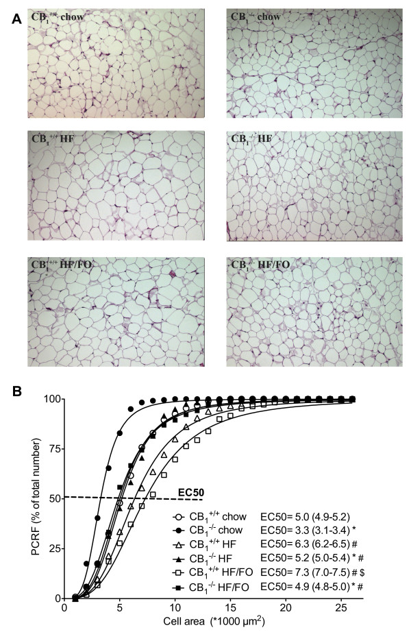
Figure 2.
Fat cell area in CB1+/+ and CB1-/- mice fed chow, a HF or a HF/FO diet during 6 weeks. A, Representative pictures of 3 μm paraffin hematoxylin and eosin-stained sections (1 cm = 100 μm) and B, percent relative cumulative frequency (PCRF) curves of 240-420 fat cell areas from adipose tissue sections. Inset: EC50 values of the PCRF curves and their 95%-confidence intervals. Open symbols, CB1+/+ mice; closed symbols, CB1-/- mice. # p < 0.05 compared to chow group of the same genotype; $ p < 0.05 compared to HF group of the same genotype, * p < 0.05 CB1-/- vs. CB1+/+ (p < 0.05 in case of no overlap between EC50 95%-confidence intervals).

