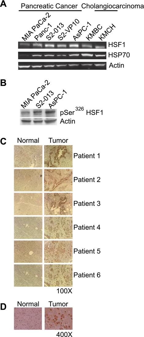
Fig. 1.
Heat shock factor 1 (HSF1) is expressed and activated in pancreatobiliary tumor cell lines and human pancreatic cancer tissue. A: representative Western blot demonstrating levels of HSF1 and heat shock protein (HSP) 70 (a transcriptional product of HSF1) in pancreatobiliary tumor cell lines. Actin was used as loading control. B: levels of HSF1 phosphorylated at serine 326 (pSer326HSF1) are higher in the more aggressive pancreatic cancer cell lines S2-013 and AsPC-1 than the less aggressive cell line MIA PaCa-2. C: HSF1 expression in pancreatic cancer tissue of 6 pancreatic cancer patients was measured by immunohistochemistry and compared with normal pancreatic tissue at the margin. Expression of HSF1 was observed to be markedly higher in pancreatic cancer tissue than normal pancreatic tissue from the margin. Negative control (no primary antibody) did not demonstrate staining (data not shown), underscoring specificity of immunostaining. D: higher-magnification images of normal and pancreatic cancer tissue from a representative patient. HSF1 staining is most prominent in the nucleus.

