
Figure 8.
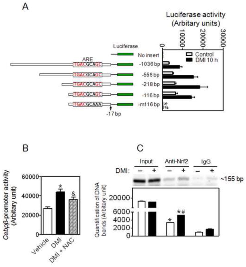
An ARE site on Cebpβ promoter is critical for its expression during adipogenesis. (A) Cebpβ promoter-reporter assay indicates an ARE site (-114∼-105 bp) in the Cebpβ promoter is critical for its expression. Left: Structures of Cebpβ promoter-luciferase reporters; Right: Activity of luciferase reporters. 3T3-L1 cells were transfected with the Cebpβ promoter-luciferase constructs when the cells reach 60-70% confluence. After 2 days confluent cells were treated with DMI for 10 h. -m116 bp, the “GC” in the ARE site were replaced with “AA”. n = 3-6; *p < 0.05 vs -116 bp Control; #p < 0.05 vs -116 bp DMI 10 h. (B) Inhibitory effect of NAC on the Cebpβ promoter activity induced by DMI. Cells expressed -116 bp Cebpβ promoter-luciferase reporter were treated with DMI with or without 5 mM NAC for 10 hrs. * p < 0.05 vs Vehicle; & p = 0.08 vs DMI alone. (C) ChIP assay indicates a physical association between Nrf2 and the Cebpβ promoter region with the ARE site. DMI, the cells were treated with DMI for 2 h. ChIP assays were performed on 3T3-L1 chromatin using either Nrf2 antibody or pre-immune antisera as negative control. Non-immunoprecipitated chromatin (1%) was used as an input control. The lower panel is the quantification of the image. n = 3; *p < 0.05 vs negative control (IgG); #p < 0.05 vs Anti-Nrf2 without DMI.