
Fig. 4.
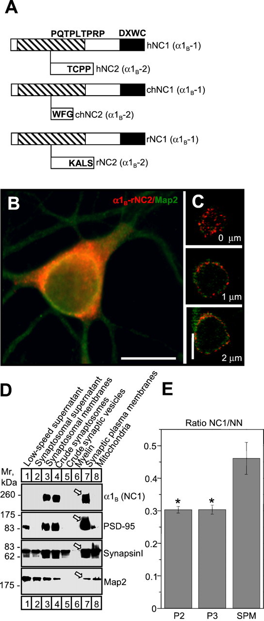
Cloning and subcellular localization of rat α1B-NC2 splice variant.A, Diagram of human (Williams et al., 1992), chicken (Lu and Dunlap, 1999), and rat α1B C-terminal splice variants. The sequence of rat α1B-NC2 has been deposited in GenBank (accession number AF389419). The position of PDZ and SH3 domain-binding motifs is indicated as in Figure 3A.B, Recombinant HA-α1B-rNC2 (red) and endogenous MAP2 (green) localization in mature neurons cultured at high density. Scale bars, 20 μm. C, Confocal analysis of recombinant HA-α1B-rNC2 localization in the soma of mature neurons cultured at low density. Three representative images from the stack are shown. D, Western blot of rat brain subcellular fractions with antibodies against rat α1B-NC1 and the neuronal markers PSD-95, synapsin I, and MAP2. Equal amounts of protein were loaded to each lane. Arrowsindicate samples on the synaptic plasma membrane fraction.E, The ratio of 125I-ω-GVIA binding site amounts precipitated by NC1 and NN antibodies from P2, P3, and SPM fractions solubilized in CHAPS. *p < 0.001.