
Fig. 8.
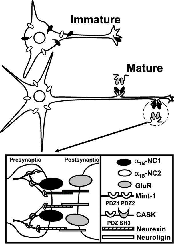
Model of N-type Ca2+ channel synaptic targeting in neurons. N-type Ca2+ channels formed by α1B-NC1 (α1B-1, CaV2.2a) (black) pore-forming subunit are distributed diffusely and uniformly in immature neurons, whereas α1B-NC2 (α1B-2, CaV2.2b) (white) is localized in the somatodendritic domain (top). After synaptogenesis (bottom), the α1B-NC1 subunits are clustered at synapses, and the α1B-NC2 subunits remain restricted to soma and dendrites. The association of α1B-NC1 C terminal with Mint1-PDZ1 and CASK-SH3 domains (Maximov et al., 1999) (inset) links synaptic N-type channels to neurexin–neuroligin neuronal adhesion complex (Irie et al., 1997; Nguyen and Sudhof, 1997; Butz et al., 1998; Song et al., 1999) and is likely to play a role in the synaptic clustering of the channels.