Figure 1.
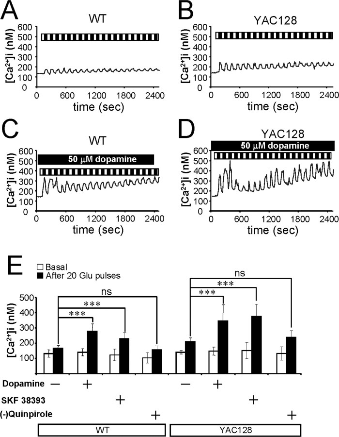
Dopamine potentiates glutamate-induced Ca2+ signals in MSNs. A, B, Repetitive application of 5 μm glutamate induces Ca2+ signals in MSNs from the WT (A) and YAC128 (B) mice. C, D, The same experiment as in A and B was performed in the presence of 50 μm dopamine with WT (C) and YAC128 (D) MSNs. Cytosolic Ca2+ levels in A–D are presented in nanomolars. Each pulse of glutamate is shown as a black bar (1 min), and the washout step is shown as a white bar (1 min) above the Ca2+ trace. The traces shown are average traces from all MSNs for each experimental group. E, The average cytosolic Ca2+ levels (mean ± SD; n is the number of MSNs analyzed) before (open bars) and after (filled bars) 20 pulses of 5 μm glutamate are shown for WT MSNs (n = 25), WT MSNs in the presence of 50 μm dopamine (n = 50), WT MSNs in the presence of 25 μm SKF38393 (n = 17), WT MSNs in the presence of 25 μm (−)-quinpirole (n = 18), YAC128 MSNs (n = 11), YAC128 MSNs in the presence of 50 μm dopamine (n = 48), YAC128 MSNs in the presence of 25 μm SKF38393 (n = 26), and YAC128 MSNs in the presence of 25 μm (−)-quinpirole (n = 13) as indicated. After 20 pulses of 5 μm glutamate, the Ca2+ levels in WT and YAC128 MSNs are significantly (***p < 0.05) higher in the presence of dopamine or SKF38393 than in its absence. In the presence of SKF38393, Ca2+ level after 20 pulses of 5 μm glutamate was significantly higher in YAC128 MSNs than in WT MSNs (p < 0.01). ns, Not significant.

