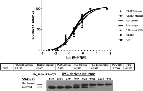
FIG. 2.
Comparison of BoNT/A1 sensitivity of hiPSC-derived neurons plated on different substrates. At 2 weeks after plating, the cells were exposed to the indicated concentrations of BoNT/A1 and cell lysates were analyzed for SNAP-25 cleavage by Western blot. Data from three Western blots were quantified by densitometry, and data plots were generated. There were no statistically significant differences in sensitivity of neurons grown on the different substrates. The substrates are shown in the figure legend on the right.

