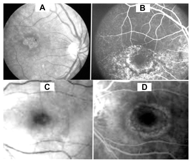
Figure 1.
(A) Fundiscopic photo of the retina showing a target maculopathy in a patient with chloroquine retinopathy. (B) Fluorescein angiography of same case showing a fairly large area of dye leakage extending from the perimacular region. (C) Fundiscopic photo of the target maculopathy in a patient with hydroxychloroquine retinal toxicity. (D) Fluorescein angiography of this case shows a milder change than seen with chloroquine.

