Figure 1.
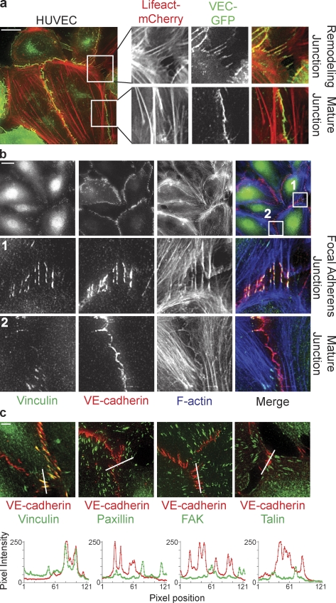
Vinculin marks distinct, remodeling cell–cell junctions attached to radial actin bundles. (a) Still images and enlarged views from time-lapse recordings (Video 2) showing perpendicularly oriented remodeling cell–cell junctions and linear stable/mature cell–cell junctions in a monolayer of HUVECs expressing VE-cadherin–GFP (green) and the F-actin probe Lifeact-mCherry (red). (b) IF images of HUVECs stained for Vinculin (green), VE-cadherin (red), and F-actin (blue) showing specific colocalization of Vinculin with perpendicular remodeling junctions, the FAJs (middle), and the absence of Vinculin from stable/mature linear junctions (bottom). (c, top) Merged IF images of HUVECs stained for Vinculin, phospho-Y118-Paxillin, phospho-Y397-FAK, or Talin (green) together with VE-cadherin (red). (c, bottom) Accompanying fluorescence intensities along the depicted lines showing that Vinculin, but not Paxillin, FAK, or Talin (green lines) colocalize with VE-cadherin (red lines) at FAJs. See also Fig. S1 for details. Bars: (a and b) 20 µm; (c) 5 µm.

