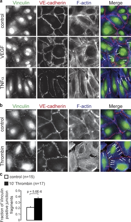
Figure 2.
VEGF, TNF, and Thrombin induce the formation of FAJs. (a) IF images of Vinculin (green), VE-cadherin (red), and F-actin (blue) in HUVEC monolayers that were left untreated (control) or stimulated with VEGF for 4 h or TNF for 24 h. Arrows point to FAJs that are characteristic for VEGF and TNF treatments. Bar, 20 µm. (b) IF images of HUVECs that were untreated or stimulated with thrombin for 10 min, and stained as in a. Please note the strong induction of FAJs (arrows) by thrombin. Bar, 20 µm. (c) Graph shows a quantification of the fraction of Vinculin-positive junction fragments (detected by automated image segmentation based on VE-cadherin signal, see Materials and methods) in control (n = 15 images) and in thrombin (n = 17 images)-treated HUVECs of two independent experiments. Values are averages ± SEM (error bars). P-value was calculated with a two-tailed, homoscedastic Student’s t test.

