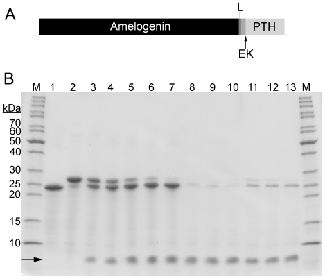
Figure 3.
A) Schematic structure of the amelogenin-PTH fusion protein. The amelogenin (in the N-terminal) and PTH (in the C-terminal) are separated by a glycine-serine linker (L) and an enterokinase site (EK). The arrow indicates the cleavage point of enterokinase. B) SDS-PAGE gel for analysis of amelogenin-PTH cleavage with enterokinase. Lane 1 is amelogenin purified with acid/heat treatment method and lanes 2–7 are purified amelogenin-PTH at different time points during enterokinase cleavage: 0 h, 1 h, 2 h, 4 h, 8 h and 24 h. Lanes 8–10 are samples after 4 h, 8 h and 24 h of cleavage where the amelogenin and amelogenin-PTH have been removed by precipitation at pH 7. Lanes 11–13 are also samples after 4 h, 8 h and 24 h of cleavage, but here the amelogenin and amelogenin-PTH have been removed by precipitation using 250 mM NaCl. Lanes marked M contain molecular weight marker. PTH is indicated with an arrow.

