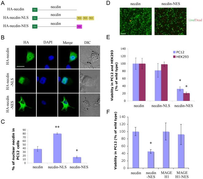
Figure 5. Exclusion of necdin from the nucleus causes cell death.
A. Schematic of necdin constructs, fused at the C-terminus to either three repeats encoding DPKKKRKV (NLS) or YLVQIFQELTL (NES). B. Representative confocal images of PC12 cells 48 hr after transfection, fixed and stained as indicated. Scale bar 10 µm. C. Quantification of nuclear localization of the indicated constructs in PC12 cells, * denotes p<0.05, ** denotes p<0.01, n = 16. D. Live/dead staining of PC12 transfected as indicated and plated on Poly-L lysine coated cover slips. 30 hours after transfection, cells were washed and incubated with 1 µM Calcein AM and 1 µM EthD-1 in D-PBS for 25 minutes, followed by fluorescence imaging. Live cells are green, dead cells are red. Scale bar 100 µm. E. Cell death in necdin-NES transfected PC12 and HEK293 cells as observed by XTT assay. F. Viability of PC12 cells transfected with the indicated constructs, 48 hours after transfection. * indicates p<0.02, n = 3.

