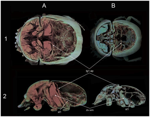
Figure 5. Micro-CT volume rendering reconstruction images of the internal anatomy.
(A) Scarabaeus sacer and (B) S. cicatricosus. Dorsal (1) and lateral (2) views showing the cuticular membrane formed dorsally by the apodeme of the first abdominal terguite, being clearly evident the bigger development of the cuticular membrane of S. sacer in comparison with that of S. cicatricosus. Abbreviations: dv-wm: dorsoventral wing muscle; st1: 1st abdominal sternite; tg1-ap: apodeme of the first abdominal terguite.

