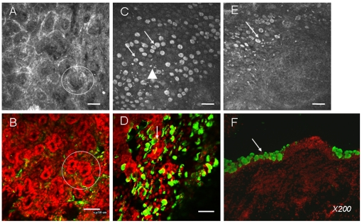
Figure 1. The morphological correlation between IVCM analysis (A, C, E) and immunohistology (B, whole-mount conjunctiva; D, impression cytology; F, cryosections) for observing the microstructure of superior conjunctiva in normal rabbits.
Bar: 50 µm. IVCM observation for meibomian acinar glands (Fig. A, circle) is similar to that of whole-mount conjunctiva with partial eyelids (Fig. B, circle). Goblet cells presented large hyperreflective, round or oval aspects (arrows in C for IVCM and D for impression cytology). Several white hyperreflective inflammatory cells were found (triangles). The CALT structure under the IVCM presented a round/oval aspect (Fig. E) with the goblet cells surrounding them. Immunohistology of MUC-5AC in cryosections confirmed this distribution of goblet cells (F).

