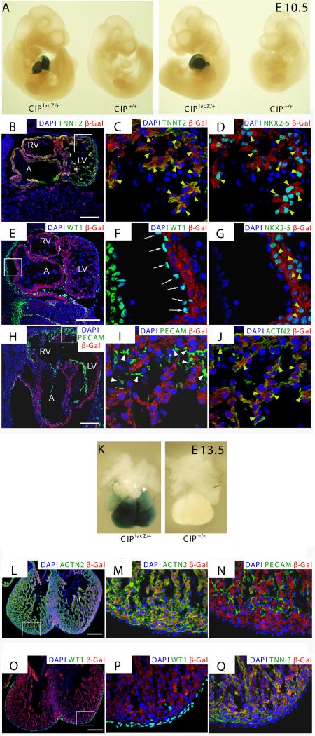
Figure 4. Characterization of CIP expression using a LacZ reporter gene in mouse embryos.
(A) Whole mount β-gal staining of mouse E10.5 embryos harboring a LacZ cassette inserted in the CIP locus (CIPlacZ/+), or the control (CIP+/+). Positive β-gal staining is only detected in the heart.
(B-J) Immunohistochemistry using indicated antibodies on tissue sections of E10.5 CIPlacZ/+ embryo. Note that CIP positive cells (marked by antibodies against the β-gal and is in red) overlay with Nkx2-5 (D, G), cardiac α-actin (ACTN2) (J) and cardiac troponin T (TNNT2) (B, C) positive cardiomyocytes (yellow arrowheads) but not that of WT1-positive epicardium (white arrows) (E, F) nor PECAM-positive fibroblasts (white arrowheads) (H, I). DAPI (blue) staining marks the nucleus.
(K) Whole mount β-gal staining of mouse E13.5 embryonic hearts harboring a LacZ cassette inserted in the CIP locus (CIPlacZ/+), or the control (CIP+/+).
(L-Q) Immunohistochemistry using indicated antibodies on tissue sections of E13.5 CIPlacZ/+ embryo. Note that CIP positive cells (marked by antibodies against the β-gal) overlay with cardiac α-actin (ACTN2) and cardiac troponin T (TNNT2) positive cardiomyocytes not that of WT1 positive epicardium, nor PECAM positive fibroblasts. DAPI staining marks the nucleus. (B, E, H, bar=100 μm; C, D, F, G, I, J, bar=10 μm; L, O, bar=200 μm, M, N, P, Q, bar=40 μm. Data are presented as representative images derived from >3 embryos. A, atrium; LV, left ventricle; RV, right ventricle.

