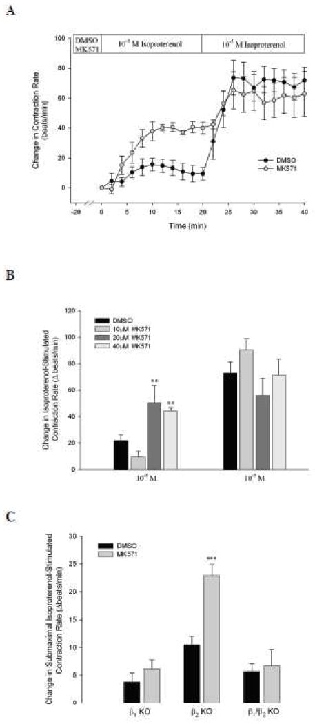
Fig. 3. Pharmacological inhibition of MRP4 by MK571 potentiates submaximal isoproterenol-stimulated contraction rate via β1-adrenoceptors.
A. WT syncytial cardiomyocytes were exposed to DMSO (n = 8) or MK571 (40 μM, n = 5) for 20 min prior to isoproterenol stimulation. Subsequently, myocytes were stimulated with 10−8 M, then 10−5 M isoproterenol for 20 min each. B. Similarly, additional experiments were carried out with 10 or 20 μM MK571 (n ≥ 5) and peak changes in contraction rates were compared to DMSO. **, P < 0.01 vs. DMSO by Student’s t-test. C. Spontaneously beating neonatal ventricular myocytes from β1-, β2-, and β1/β2 KO mice were exposed to DMSO (n ≥ 6) or MK571 (40 μM, n ≥ 6) in a similar manner as above with peak responses to submaximal isoproterenol stimulation (10−8 M) represented. ***, P < 0.001 vs. DMSO for each group by Student’s t-test.

