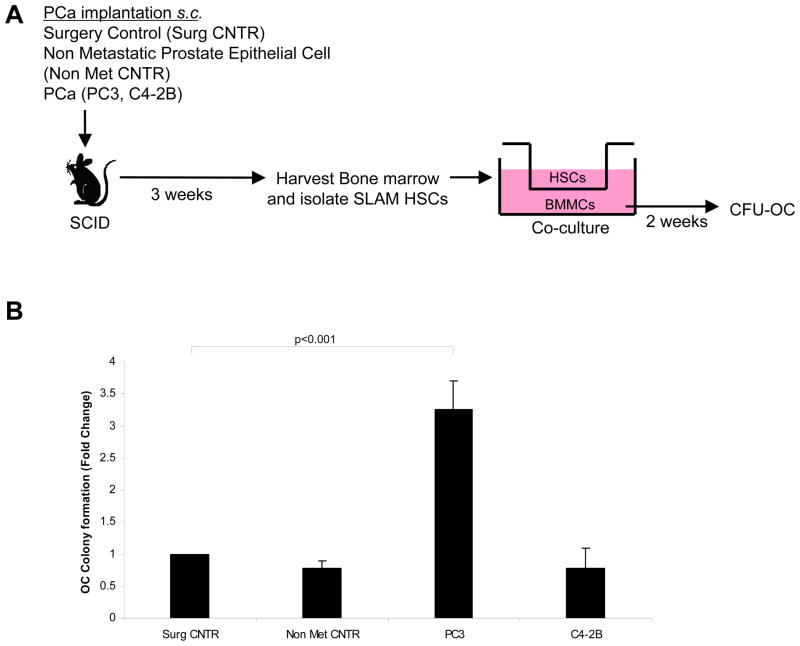
Figure 3. HSCs from osteolytic PCa implanted animals induce osteoclastic colony forming units from BMMCs.
(A) Experimental outline. Co-cultures were established by placing SLAM HSCs (CD150+ Lin− CD48− CD41− Sca-1+ cKit+) derived from animals bearing s.c. tumors or control animals in the top chamber of a dual culture well plate, and mixed mononuclear bone marrow cells in the bottom well. After 2 weeks the phenotype of the resulting colonies were stained for TRAP to visualize osteoclast colonies (CFU-OC). (B) Number of TRAP stained cells/culture. Significant increase in CFU-OCs was noted in the presence of HSCs derived from PC3 tumor-bearing animals vs. surgical control animals. Data represent mean ± SD fold change performed in triplicate in 3 independent cultures. Significance (p<0.001) were calculated compared to surgical control treated group.

