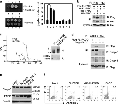
Figure 6.
Truncated FADD (tFADD) exhibits enhanced self-association to recruit procaspase-8 resulting in casp-8 activation. (a) tFADD exerts stronger self-association than FL-FADD. AH109 yeast strain was co-transformed with the indicated constructs and the transformants were plated on synthetic defined medium with or without histidine (−His) and adenine (−Ade). −His−Ade plates were supplemented with 1.5 mM 3-AT to suppress background growth. +His+Ade plates were served as identical amount controls of yeasts. AH109 cotransformed with pGADT7-SV40 large T-antigen and pGBKT7-murine p53 was used as a positive control (left panel). Interaction activity was quantified by β-galactosidase assay and shown as means±S.D. (right panel). **P<0.01. 1: AD-FL-FADD+BD-FL-FADD; 2: AD-tFADD+BD-tFADD; 3: AD-SV40 large T antigen+BD-murine p53; 4: AD vector+BD vector; 5: AD-FL-FADD+BD-murine p53; 6: AD-SV40 large T antigen+BD-FL-FADD; 7: AD-tFADD+BD-murine p53; 8: AD-SV40 large T antigen+BD-tFADD. (b) Self-association of tFADD was confirmed by Co-IP assay. Flag-FL-FADD and HA-FL-FADD or Flag-tFADD and HA-tFADD were cotransfected into HEK293A cells. After 24 h, cells were harvested and lysed for immunoprecipitation with anti-Flag antibody and visualized by immunoblotting with anti-HA and anti-Flag antibodies. Mouse IgG was used as a negative control. (c) tFADD facilitates its oligomerization. GST-FL-FADD or GST-tFADD recombinant proteins were analyzed by gel filtration (Superdex 200 column) (left panel). GST-FL-FADD or GST-tFADD was observed on reduced (R, lower panel) and native (N, upper panel) gels. D, dimmer; M, monomer; P, polymer. (d) tFADD efficiently recruits procaspase-8. HEK293A cells were transfected with Flag-tagged FL-FADD or tFADD for 24 h followed by casp-8 immunoprecipitation. (e) Overexpression of tFADD causes casp-8 activation. HEK293A cells were infected with lentivirus encoding FL-FADD, GFP, M196A-FADD or tFADD for 24 h followed by immunoblotting. The same blot was stripped for western blotting. (f) tFADD overexpression causes apoptosis. Cells overexpressing with the indicated proteins for 24 h were analyzed by Annexin V/PI staining. The data are representative of at least three independent experiments

