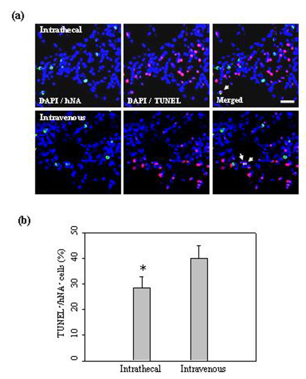
Figure 2.
hUCB-MSCs undergoing apoptotic cell death in the ischemic brain. (a) At seven days after 1 × 10⁶ hUCB-MSC administration, hUCB-MSCs undergoing apoptotic cell death were measured by TUNEL staining (n = 5 per treatment group). hUCB-MSCs were identified by the staining with human nuclei antibody (hNA, green). The numbers of hNA-TUNEL double-positive cells in the ipsilateral ischemic boundary zone (IBZ) are illustrated. (b) Quantitative analysis of hNA-TUNEL double-positive cells in the ipsilateral IBZ. Data from five animals are presented as mean values ± SD. There were significantly more hNA-TUNEL double-positive cells in animals after intravenous administration (analysis of variance; *P < 0.05). Nuclei were counterstained with DAPI (blue). Scale bar = 20 μm.

