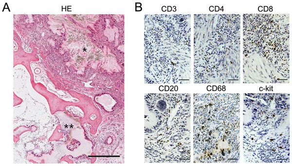
Figure 1.
Infiltration of chronic inflammatory cells in gouty tophus tissues. (a) Tophus tissue sections from patients with chronic gouty arthritis were processed for hematoxylin-eosin (HE) staining. * indicates monosodium urate monohydrate (MSU) crystals, and ** represents osteolytic lesions. (b) Serial sections were processed for immunohistochemical staining using anti-CD3, anti-CD4, anti-CD8, anti-CD20, anti-CD68, and anti-c-kit antibodies (brown). Scale bar in a = 500 μm; scale bars in b = 50 μm.

