Figure 3.

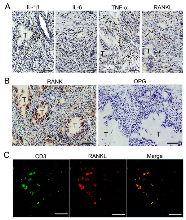
Expression of pro-resorptive cytokines in gouty tophus tissues. (a, b) Tophus tissue sections from patients with chronic gouty arthritis were processed for immunohistochemical staining using anti-interleukin (IL)-1β, anti-IL-6, anti-tumor necrosis factor (TNF)-α, anti-receptor activator of nuclear factor κB (RANK), anti-osteoprotegerin (OPG), and anti-receptor activator of nuclear factor κB ligand (RANKL) antibodies (brown). T, gouty tophus. (c) Immunofluorescent stains for CD3 (green) and RANKL (red) in tophaceous gout tissues. Scale bars in a, b, and c = 50 μm.
