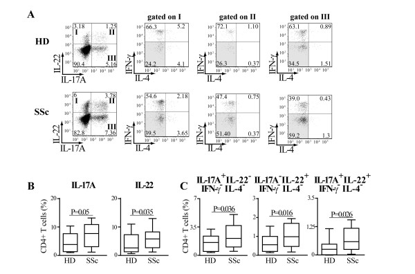
Figure 2.
Th17 and Th22 cells are preferentially expanded in SSc individuals. PBMC were activated by CD3/CD28 crosslinking and cultured in the presence of IL-2 (20 U/ml). A. FACS plots gated on CD4+ T cells of cells harvested at Day 7 of culture and activated by PMA/ionomycin for a representative HD and SSc individual. Intracellular cytokine accumulation was detected by five-color flow cytometry. Numbers in plots indicate the percentage of cells in each quadrant. IFN-γ and IL-4 double negative cells are highlighted by grey shading. B. Frequency of IL-17A+ and IL-22+ CD4+ T cells in 29 HD and 30 SSc individuals detected by flow cytometry. C. Frequency of CD4+ T cells producing IL-17A alone (IL-17A+IL-22-IFN-γ-IL-4-cells), IL-22 alone (IL-17A-IL-22-IFN-γ-IL-4-cells), and IL-17A in combination with IL-22 (IL-17A-IL-22-IFN-γ-IL-4-cells) in 24 HD and 29 SSc individuals detected by multiparameter flow cytometry analysis.

