
Figure 7. Histopathologic assessment of lung tissue.
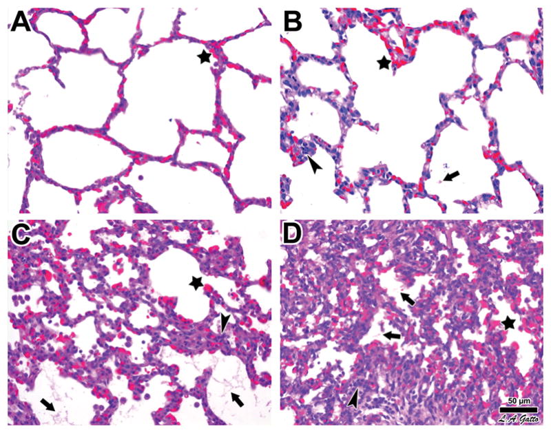
Swine were subjected to endotoxemia and survivor animals sacrificed at the end of the experimental period. Upon death, their lungs were processed for histology and hematoxylin & eosin staining as described in the Materials and Methods. Panel A. The lungs of Pig 2588 demonstrated normal histology except for blood vessel congestion conspicuous at all levels, including alveolar capillaries (star). Panel B. The lungs of Pig 2595 exhibited a higher level of histological involvement marked by the focal presence of fibrinous deposits in the alveolar lumen (arrow), along with greater infiltration of leukocytes (arrowhead), in addition to alveolar capillary congestion (star). Panel C. The lungs of Pig 2583 showed all signs of ARDS, including alveolar capillary congestion (star), fibrinous deposits (arrows), and abundant leukocyte infiltration (arrowhead). Panel D. The lungs of Pig 2589 showed the most pronounced signs of acute lung injury, with widespread alveolar capillary congestion (star), fibrinous deposits in the air compartment (arrows), and marked cellularity associated with more severe leukocyte infiltration (arrowhead). The progression from A to D is also marked by increased atelectasis. Bar = 50 μm.