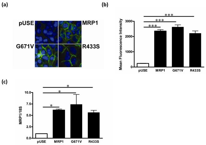
Fig. 1. Expression in stable cell lines of multidrug resistance-associated protein 1 (MRP1) and is variants.
(a) Cells were immunostained for MRP1 protein; blue = nuclei, green = MRP1 protein. Plasmids containing wild-type MRP1 or the MRP1 variants G761V and R433S were transfected into HEK293 cells to generate stable cell lines. pUSE cells were HEK293 cells transfected with pUSEamp(+) empty vector and used as a control. (b) Cells were stained with MRPr1 primary antibody, followed by fluorescence labeled secondary antibody (AlexaFlor488) for flow cytometry analyses. Bar graphs show mean fluorescence intensity ± S.E. (c) MRP1 mRNA expression was detected by real time RT-PCR and normalized by 18S rRNA expression. * P < 0.05, *** P < 0.001.

