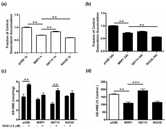
Fig. 3. DOX and GS-HNE retention in HEKpUSE, HEKMRP1, HEKR433S, and HEKG671V cells.
(a) Cells were cultured in the presence or absence of 50 μM DOX for 1 h, followed by a 30 min efflux period, and analyzed for intracellular DOX by fluorescence spectrophotometry. (b) Cells were cultured in media alone or in the presence of DOX (1.5 μM) for 24 h. Cells were harvested, lysed and intracellular concentrations of DOX (c) and GS-HNE (d) determined. (d) The concentration of GS-HNE in cells expressed as a percentage of that in HEKMRP1 cells. ** P < 0.01, *** P < 0.001

