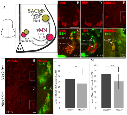
Fig. 2.
Appropriate numbers of properly specified SACMNs are formed in Nkx2.9-deficient mice. (A) Schematic of SACMN and ventral motor neuron (vMN) development in the ventral spinal cord. SACMNs (green circle) and vMNs (red circle) arise from MN progenitors, which migrate away from the ventricular zone to settle in dorsolateral or ventrolateral regions, respectively, of the spinal cord. (B-G) Cryosections derived from WT (B-E, E10.5; F,G, E11) embryos labeled with various SACMN or vMN markers show that SACMNs and their axons (arrow) migrate through vMNs [SACMN (v)] and SACMNs that have settled near the LEP [SACMN (d)] and express BEN, Islet1 and Phox2b, whereas vMNs express Islet1 and HB9. C, E and G are magnified views of the boxed areas in B, D and F, respectively. (H-K) E11.5 WT (H,I) and Nkx2.9–/– (J,K) embryos were labeled with anti-Phox2b (H,J) and colabeled with anti-Phox2b and anti-BEN (I,K). I and K are magnified views of the boxed areas in H and J, respectively. (L,M) The number of Phox2b+ BEN+ (E11.5; n=4) and Islet1+ BEN+ (E10.5; n=3) SACMNs is not statistically different between Nkx2.9–/– and WT/heterozygote littermates. Error bars indicate s.d. Scale bars: 50 μm in F for B,D,F, in J for H,J; 25 μm in G for C,E,G, in K for I,K.

