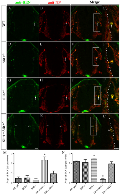
Fig. 6.
SACMN axon exit is perturbed in Slit1–/–Slit2–/– mice. (A-L′) Cryosections derived from E10.5 WT (A-C′) and Slit mutant (D-L′) embryos were labeled with anti-NF and anti-BEN. SACMN axons appropriately exit the spinal cord and assemble into an external SAN (arrow) in WT (A-C′; E10.5, n>5; E11.5, n>5), Slit1–/– (D-F′; E10.5, n=5; E11.5, n=9; data not shown), Slit2–/– (G-I′; E10.5, n=1; E11.5, n=3) or Slit1–/– Slit3–/– (E11.5, n=3; data not shown) mice. (M,N) The average number of SANs located inside or outside of the spinal cord is not statistically different between Slit1–/– mutant, Slit1–/– Slit3–/– mutant and WT mice. Notably, in Slit1–/– Slit2–/– mice, SACMN axons fail to exit the CNS and instead assemble into an ectopic SAN (J-L′, asterisks). The average number of SANs outside the spinal cord in Slit2–/– mice is significantly increased compared with WT embryos (N). *P<0.05; error bars indicate s.e.m. E10.5, n=3; E11.5, n=3; E11.5 data not shown. Scale bars: 50 μm in J for A-C,D-F,G-I,J-L; 25 μm in L′ for C′,F′,I′,L′.

