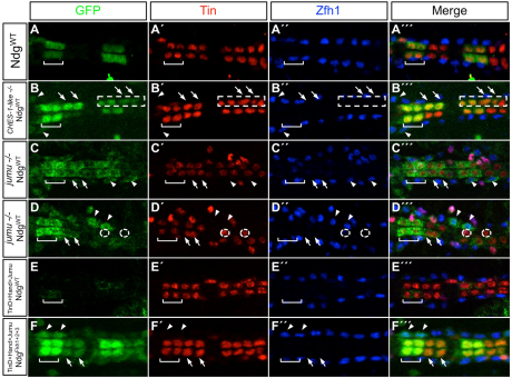
Fig. 5.
Jumu and CHES-1-like repress activity of the Ndg enhancer in the heart. (A-A″′) A GFP reporter driven by the wild-type Ndg enhancer is expressed in only two cardial cells per hemisegment, the Tin-Lb-CCs (square bracket). (B-B″′) Reporter expression from the wild-type Ndg enhancer is de-repressed into additional CCs (arrows) and Odd-PCs (arrowheads) in embryos homozygous for the CHES-1-like null mutation. The image shows de-repression occurring in both morphologically normal and abnormal (boxed) hemisegments. (C-D″′) Similar de-repression into additional CCs (arrows) and Odd-PCs (arrowheads) is seen for both normal (C-C″′) and abnormal (D-D″′) hemisegments in embryos homozygous for the jumu-null deficiency. The dotted circles in D-D″′ outline the two (instead of four normal) Tin-expressing CCs present in an abnormal hemisegment. (E-E″′) Overexpression of Jumu in the cardiac mesoderm and heart represses reporter expression from the wild-type Ndg enhancer, even in the two Tin-Lb-CCs (square bracket) where Ndg is normally expressed. (F-F″′) Overexpression of Jumu is unable to repress reporter expression from an enhancer with mutations in all three Fkh sites. Rather, these embryos exhibit de-repression into additional CCs (arrows) and Odd-PCs (arrowheads), similar to that seen with wild-type levels of jumu expression (Fig. 4F-F″′). Abdominal segments A3 and A4 are shown in every panel.

